Human Leg Bones Diagram - Practical Art Anatomy E G Lutz - See more ideas about muscle anatomy, human anatomy and physiology, body anatomy.
byAdmin-
0
Human Leg Bones Diagram - Practical Art Anatomy E G Lutz - See more ideas about muscle anatomy, human anatomy and physiology, body anatomy.. The bones of the leg and foot form part of the appendicular skeleton that supports the many muscles of the lower limbs. Browse 7,069 leg bone stock photos and images available, or search for human leg bone or leg bone xray to find more great stock photos and pictures. Human anatomy for muscle, reproductive, and skeleton. At the same time, the bones and joints of the leg and foot must be strong enough to support the body's weight while remaining. Also called the shin bone, the tibia is the longer of the two bones in the.
Distal end of right humerus. It is controlled by the obturator nerve. Its decrease finish helps create the knee joint. They allow you to move and provide support for your upper body. File human arm bones diagram svg wikipedia.
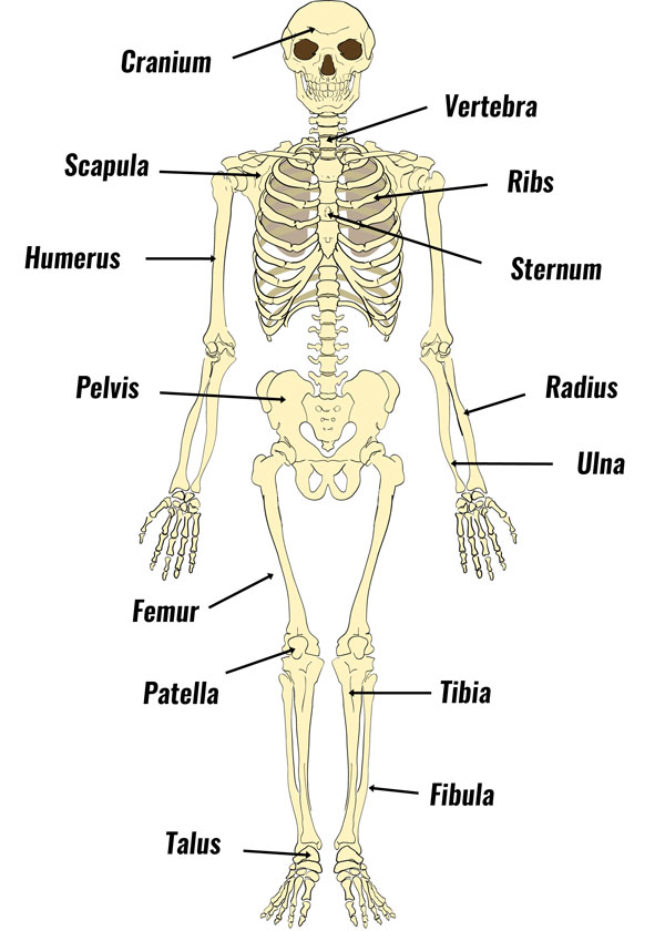
Leg Bone Anatomy Diagram Diagram Of Human Leg Human Anatomy Diagram from www.anatomynote.com You'll learn about the muscle. Browse 7,069 leg bone stock photos and images available, or search for human leg bone or leg bone xray to find more great stock photos and pictures. At the same time, the bones and joints of the leg and foot must be strong enough to support the body's weight while remaining. Related posts of diagram of leg bones. The human leg, in the general word sense, is the entire lower limb of the human body, including the foot, thigh and even the hip or gluteal region. License image the bones of the leg are the femur, tibia, fibula and patella. Together, the upper and lower legs and the feet make up half the length of the human figure. The bones of the leg are the femur, tibia, fibula and patella.
The human leg, in the general word sense, is the entire lower limb of the human body, including the foot, thigh and even the hip or gluteal region.
At the same time, the bones and joints of the leg and foot must be strong enough to support the body's weight while remaining. The knee joint is the largest joint in the body and is primarily a hinge joint, although some sliding and rotation occur. See more ideas about muscle anatomy, human anatomy and physiology, body anatomy. Leg muscle sport trauma and bone pain labeled diagram. The human leg, in the general word sense, is the entire lower limb of the human body, including the foot, thigh and even the hip or gluteal region. Together, the upper and lower legs and the feet make up half the length of the human figure. A high ankle sprain causes pain and swelling similar to a. Related posts of muscles and tendons of the leg muscle anatomy head. File human arm bones diagram svg wikipedia. The bones of the leg are the femur, tibia, fibula and patella. The human leg consists of 8 bones, 4 per leg. The bones of the leg and foot form part of the appendicular skeleton that supports the many muscles of the lower limbs. The largest and most medial leg bone, forming both the knee and ankle joints.
The knee joint is the largest joint in the body and is primarily a hinge joint, although some sliding and rotation occur. Human leg bones diagram : The ligament joining the two bones of the lower leg (tibia and fibula), called the syndesmotic ligament, is injured. Legs are used for standing, and all forms of. This diagram depicts human leg bone anatomy.human anatomy diagrams show internal organs, cells, systems, conditions, symptoms and sickness information and/or tips for healthy living.
The Human Skeleton Bones Structure Function Teachpe Com from www.teachpe.com File human arm bones diagram svg wikipedia. 12 photos of the bones leg diagram picture. The femur is the largest bone in the body and the only bone of the thigh (femoral) region. The bones of the leg are the femur, tibia, fibula and patella.the foot bones shown in this diagram are the talus, navicular, cuneiform, cuboid, metatarsals and calcaneus. The bones of the leg are the femur, tibia, fibula and patella. The bones together make up the hip. This diagram depicts diagram leg bones anatomy.human anatomy diagrams show internal organs, cells, systems, conditions, symptoms and sickness information and/or tips for healthy living. Formed by the left and right hip bones, the pelvic girdle connects the lower limb (leg) bones to the axial skeleton.
This area is commonly referred to as the calf.
The human leg, in the general word sense, is the entire lower limb of the human body, including the foot, thigh and even the hip or gluteal region. Distal end of right humerus. File human arm bones diagram svg wikipedia. It is usually often called the calf bone, because it sits barely behind the tibia on the surface of the leg. Leg muscle sport trauma and bone pain labeled diagram. Formed by the left and right hip bones, the pelvic girdle connects the lower limb (leg) bones to the axial skeleton. The bones of the hip include the femur, the ilium, the ischium, and the pubis. The bones together make up the hip. Human anatomy diagrams show internal organs, cells, systems, conditions, symptoms and sickness. The knee joint is the largest joint in the body and is primarily a hinge joint. Lacrimal bone diagram wiring diagram. The bones of the leg are the femur, tibia, fibula and patella.the foot bones shown in this diagram are the talus, navicular, cuneiform, cuboid, metatarsals and calcaneus. They allow you to move and provide support for your upper body.
It is controlled by the obturator nerve. The human leg consists of 8 bones, 4 per leg. The knee joint is the largest joint in the body and is primarily a hinge joint. Master leg and knee anatomy using our topic page. Diagram of leg bones, find out more about diagram of leg bones.
Home Anatomy Physiology For Ems Libguides At Com Library from s-media-cache-ak0.pinimg.com You'll learn about the muscle. Related posts of muscles and tendons of the leg muscle anatomy head. Its lower end helps create the knee joint. Browse 7,069 leg bone stock photos and images available, or search for human leg bone or leg bone xray to find more great stock photos and pictures. Its decrease finish helps create the knee joint. The hip itself is a ball and socket joint, much like the shoulder.the structures necessary to create this joint are the socket, the joint capsule, muscle, ligaments, and the neck. The ligament joining the two bones of the lower leg (tibia and fibula), called the syndesmotic ligament, is injured. The largest and most medial leg bone, forming both the knee and ankle joints.
Each leg is made up of four bones.
Its lower end helps create the knee joint. The pubis, ischium, and ilium together constitute the pelvis while the thigh bone is the femur. File human arm bones diagram svg wikipedia. Diagram of leg bones, find out more about diagram of leg bones. This diagram depicts diagram leg bones anatomy.human anatomy diagrams show internal organs, cells, systems, conditions, symptoms and sickness information and/or tips for healthy living. The femur is the largest bone in the body and the only bone of the thigh (femoral) region. The ligament joining the two bones of the lower leg (tibia and fibula), called the syndesmotic ligament, is injured. The human leg, in the general word sense, is the entire lower limb of the human body, including the foot, thigh and even the hip or gluteal region. The bones together make up the hip. It is controlled by the obturator nerve. Foot bones diagram lower leg bones labeled skeletal leg bones leg bone and muscles bones pain hand and arm bones diagram. These muscles work together to produce movements such as standing, walking, running, and jumping. Distal end of right humerus.